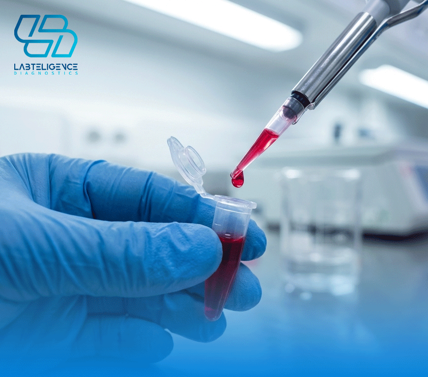
Diagnostic kits for infectious diseases in small animals are among the most widely used tools in veterinary clinical practice and internal medicine. One of the unique advantages of these kits is their rapid results compared to other laboratory methods. Although PCR is considered the gold standard for diagnosing infectious diseases such as parvovirus, due to the time required and associated costs, it is not always feasible for all suspected cases. Therefore, clinical diagnostic methods and immunochromatographic kits—classified as rapid diagnostic tools due to their complex structure—are commonly used as routine methods.
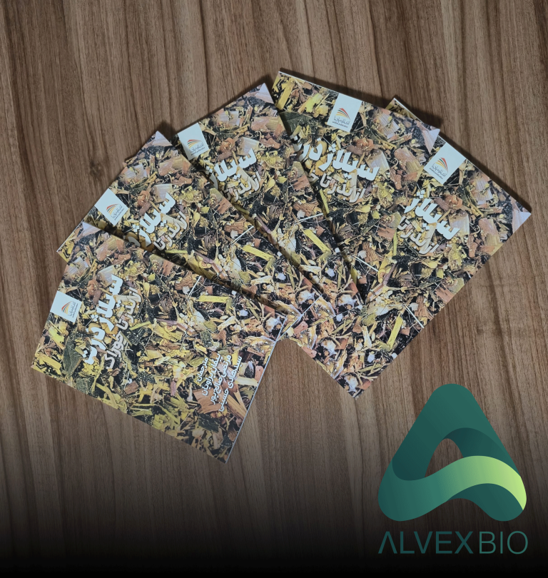
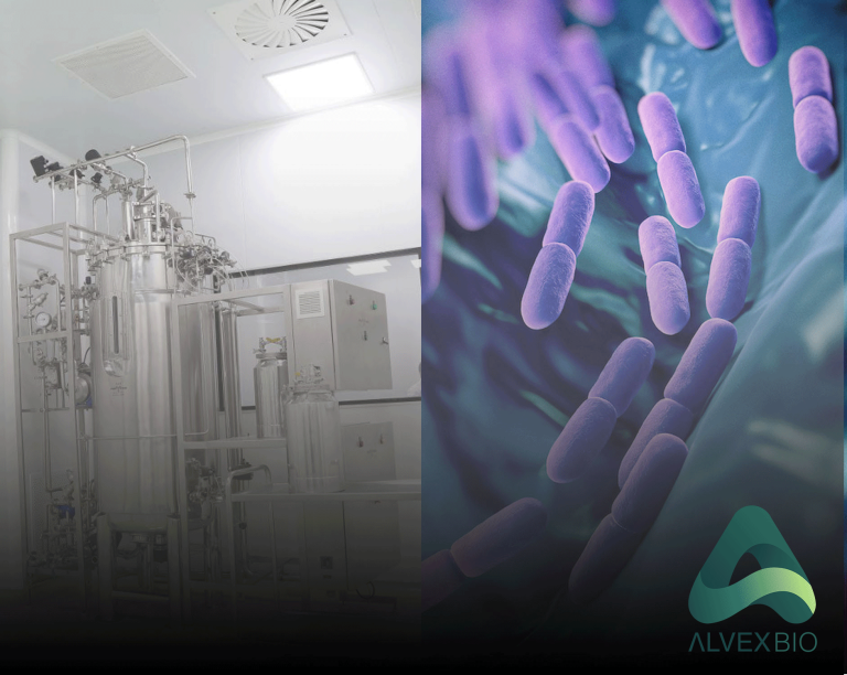
Sarshar Group is proud to announce that it has taken a significant step toward the production of veterinary diagnostic kits for the first time in the country. The group has successfully transformed complex macromolecules into practical clinical products independently, without relying on ready-made products from major global manufacturers.
Immunochromatographic assay kits, while considered fast and cost-effective solutions, have a complex structure. The Immunochromatographic Assay (also known as Lateral Flow Assay) is a rapid, simple diagnostic method based on specific antigen–antibody reactions. It is used for the qualitative or semi-quantitative detection of pathogens, hormones, toxins, or biomarkers in biological samples. Due to their speed, lack of need for advanced equipment, and point-of-care usability, these kits are widely applied in medicine, veterinary science, and the food industry.
Structurally, an immunochromatographic kit typically consists of a multilayer strip, with each layer serving a specific function in the testing process. The Sample Pad, located at the beginning, is where the sample is applied and is responsible for regulating flow and partially filtering the sample. Next is the Conjugate Pad, which contains specific antibodies bound to labeled particles (such as gold nanoparticles or colored latex). Following this is the Nitrocellulose Membrane, where the Test Line and Control Line are immobilized. At the end of the strip is the Absorbent Pad, which ensures consistent fluid flow through capillary action.
The working principle of the kit is based on capillary movement of the sample along the strip. After adding a liquid sample (such as blood, serum, urine, or extracted swab), it migrates along the strip and reaches the conjugate pad. If the target analyte (such as a specific antigen or antibody) is present, it binds specifically to the labeled antibodies, forming an immune complex. This complex continues to move with the fluid toward the nitrocellulose membrane.
At the Test Line, another antibody or antigen is immobilized, capturing the immune complex. The accumulation of labeled particles at this site produces a visible colored line, indicating a positive result. The Control Line functions independently of the analyte and captures excess labeled antibodies to confirm proper test performance and sample flow. Therefore, the appearance of the control line is essential for test validity.
Scientifically, these kits rely on key immunological properties such as high specificity, antigen–antibody affinity, and non-covalent interactions. The precise design of antibodies, type of label, quality of the nitrocellulose membrane, and capillary flow conditions all play crucial roles in determining sensitivity, specificity, and reproducibility. For this reason, quality control of raw materials and optimization of each component are critical steps in the production of immunochromatographic kits.